In some cases the bone destruction (lysis) can be extensive resulting in very significant loss of host bone stock. This can present major challenges when it comes to revision (re-do)surgery for the failed joint replacement as there may be not enough bone left to implant another joint replacement.
Unfortunately if bone loss (lysis) is very extensive it may still not be possible to restore enough bone stock for adequate fixation of the revision implants.
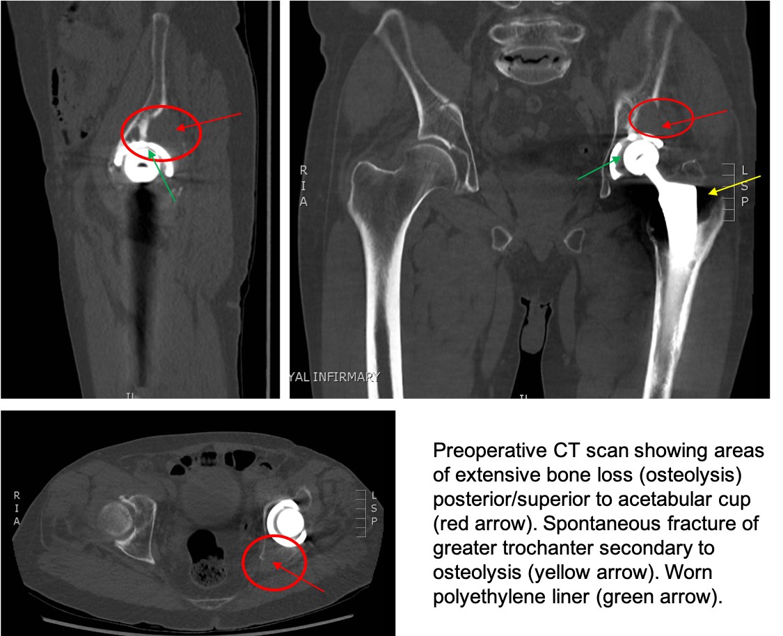
If the x-rays taken prior to planning the revision surgery suggest this may be the case, a CT scan of the hip joint is performed. This allows detailed information about the site and size of the bone loss to be calculated.
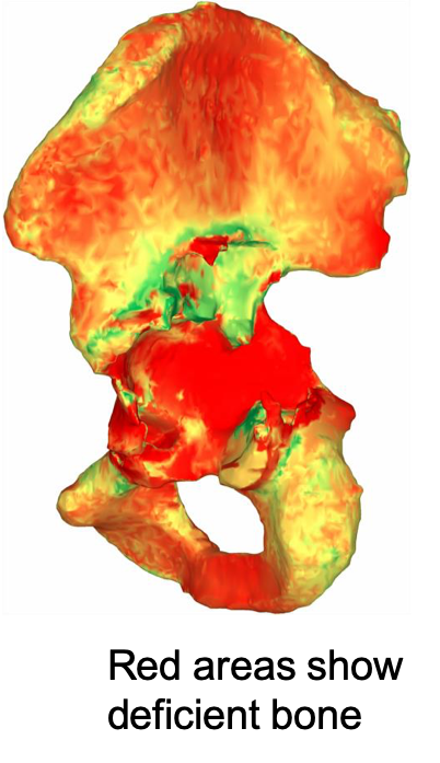
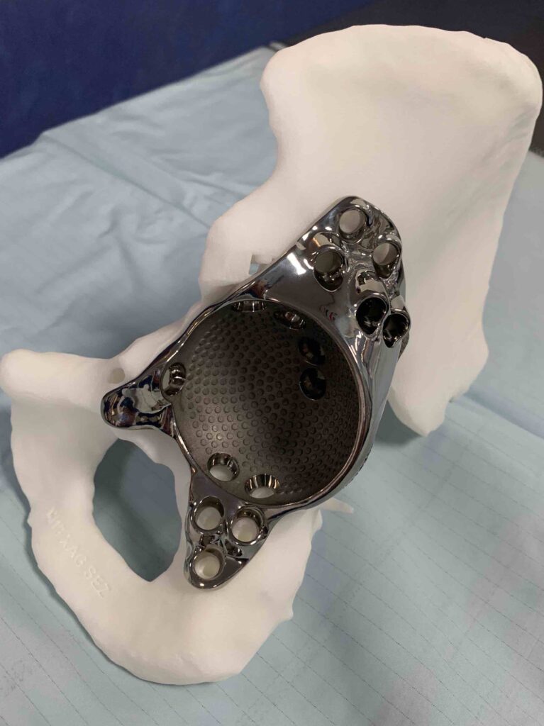
By using specialist software an assessment about the quantity and quality of the remaining bone stock for fixation of a bespoke titanium 3D printed acetabular implant that exactly matches the bone defect in the pelvis can be determined.
The 3D printed titanium implant can then be securely fixed to the pelvis using multiple screws, the direction and length of each predetermined using information derived from the planning CT scan. Custom jigs are also manufactured to direct the screws in the correct orientation to minimise the risk of injury to important pelvic structures such as major blood vessels and nerves.
Once the 3D printed pelvic socket (acetabulum) has been securely fixed to the pelvis the cup of the hip replacement can then be cemented into place in the desired orientation. The size of which is also determined by preoperative planning.
Although this is very challenging surgery, this state-of-the art technology allows revision surgery to take place that otherwise would not have been possible, thereby restoring patients function and helping maintain their quality of life.
Case example.
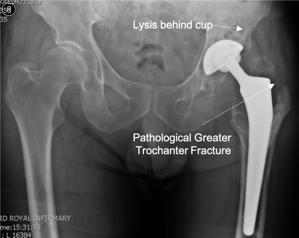
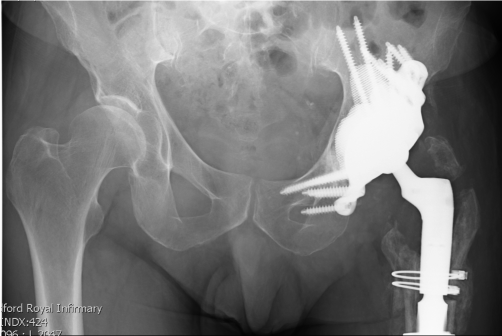
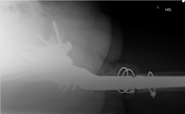
An 85 yrs. old very active gentleman underwent an uncemented THR in 2004 and presented with pain in his hip following a spontaneous fracture of his greater trochanter. He was having to use crutches for the pain. There was no history of any injury to the hip.
X-rays confirmed a pathological periprosthetic fracture of the greater trochanter. Due to concerns about significant bone loss as a result of excessive wear debris a CT scan was performed.
This confirmed extensive osteolysis behind the acetabular cup and proximal femur presenting a significant challenge for revision surgery.
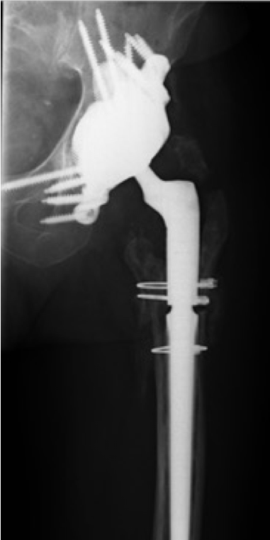
Preoperative planning confirmed the extend of the bone loss and a bespoke titanium acetabular insert was 3D printed to fit the remaining bone stock of his pelvis.
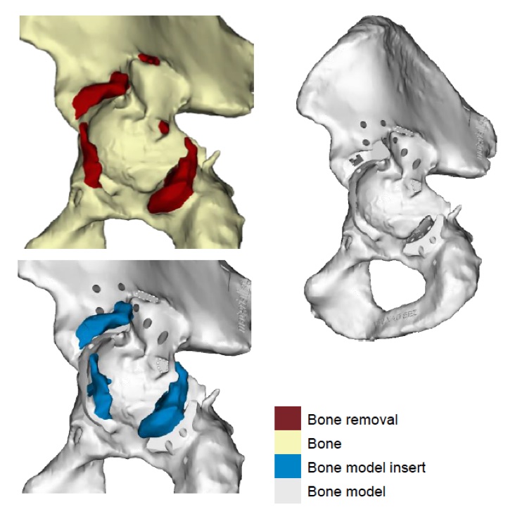
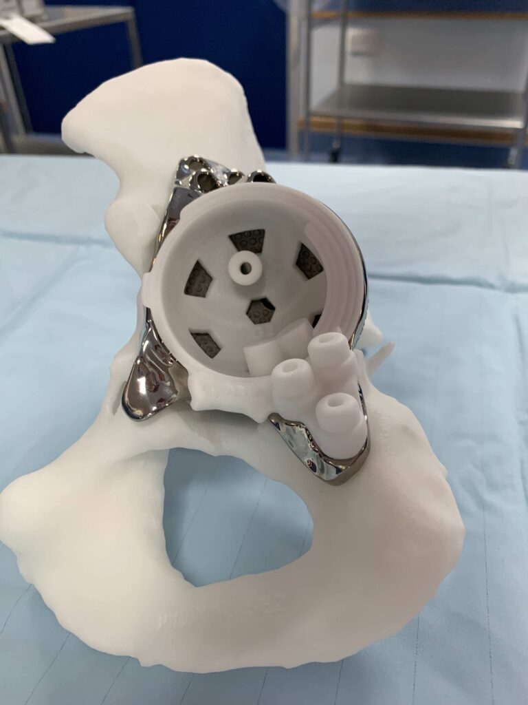
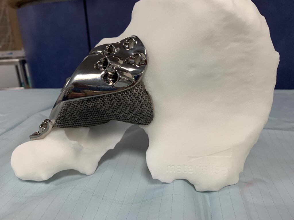
A 3D model of the patient’s pelvis as well as a model of the definitive implant are also made to allow pre and intra-operative trial fitting. Often some bone needs to be removed for access to allow the implant to be fitted properly to the pelvis and this can be visualised and practiced more effectively by using 3D models.
The bone deficient proximal femur was bypassed by using a long distally fixed uncemented stem.
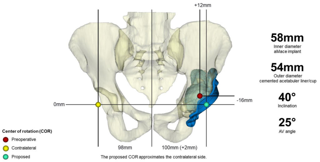
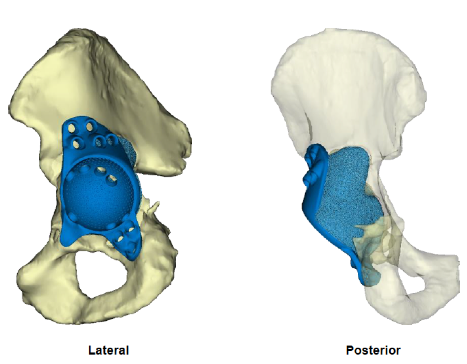
Pre-operative CT Planning
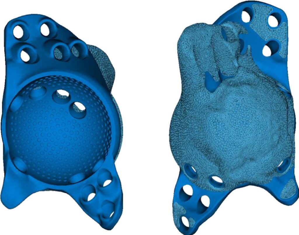
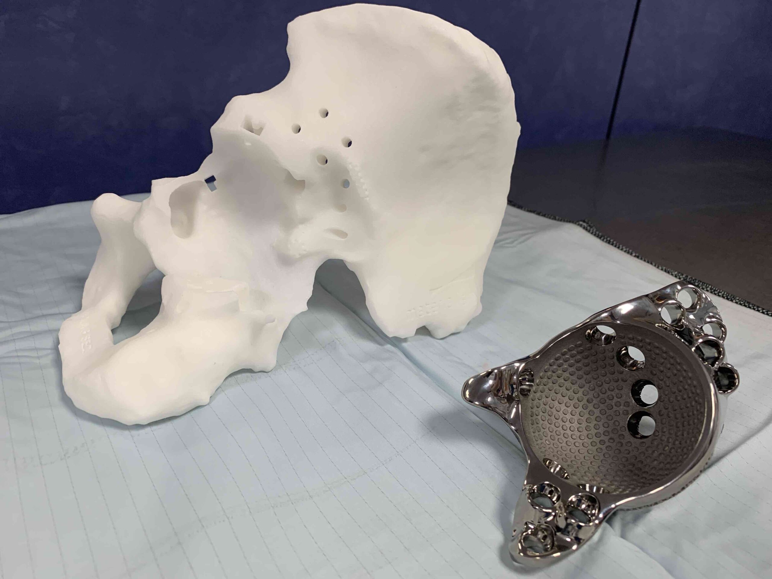
3D Printing Plans
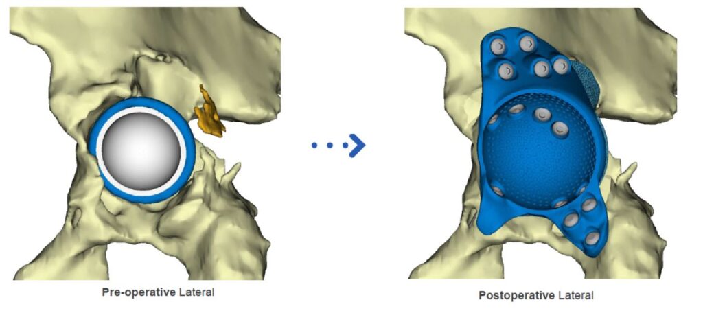
Images showing areas of bone loss in postero-superior and medial aspects of the acetabulum. Some bone has to be removed (brown) to allow bespoke 3D titanium implant to be fitted to the pelvis.
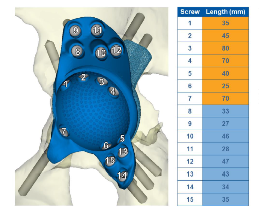
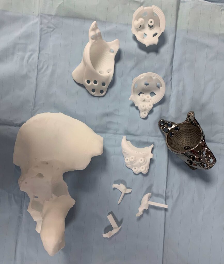
Intra-operative planning guides
Intra-operative guides showing the length, position and direction of the bone screws.
3D Printing Plastic Models of Pelvis allow pre- and intra- operative implant trialling.
3D printed screw guides and implant trials.
3D printed replica of the pelvis and implant includes the pieces of bone that need to be removed to allow exact fitting of the implant to the pelvis (bottom left of picture).